Biology concepts explained in this episode:
- What does a reduced amplitude in the ECG reading mean?
- What is an Echocardiogram?
- What is Pericardial Effusion?
- Let’s Take a Step Back: The Venous Drainage From the Head
Continuing on from the previous episode, we dig into the next events that occurred from the Pilot Episode of The Good Doctor, Season 1.
The boy who got into an accident at the airport is now being transported to St. Bonaventure Hospital in an ambulance. While inside the vehicle, Dr. Murphy claims to notice a change in the boy’s ECG which the paramedic is quick to disagree saying it has stayed the same. Dr. Murphy insists that the amplitude became lower implying a lower voltage.
What does a reduced amplitude in the ECG reading mean?
The Electrocardiogram shows the electrical activity of the heart. If something is stopping the heart from being able to fully contract, the amplitude in the ECG is reduced due to weaker signals hence, the resulting lower voltage from the heart. Dr. Murphy noticed this. He knew that there was something wrong with the boy’s heart.

As soon as they arrived at the hospital, Dr. Murphy insisted that the patient had to have an echocardiogram included as one of the first tests to be done. He made sure to relay this to the physician on duty, who then, was Dr. Claire Browne.
Dr. Browne, after having read the boy’s condition and history provided by the paramedic, believed that the boy’s cardiac results were within normal range implying that his heart was fine and that an echo was not necessary.
But, even when she believed this, somehow, Dr. Murphy’s persistence influenced her of suggesting the procedure while doing the operation on the boy with her colleagues.
What is an Echocardiogram?
An echocardiogram (ECG) is an ultrasound of the heart that helps in visualizing the organ to see if there are any structural abnormalities.
Meanwhile, inside the operating room, Dr. Browne and Dr. Neil Melendez, the attending surgeon were doing some procedures on the boy when Dr. Melendez noticed something different from the boy’s ECG.
He remembered how Dr. Browne had suggested earlier doing an echo and tried to ask why.
Dr. Browne couldn’t explain as well and only mentioned that some weird guy who did the first aid earlier at the airport suggested doing the test on him.
Dr. Melendez quickly decided to do an echo on the patient while making sure to speak with Dr. Murphy about what he knew of the patient’s condition.
Why was Echocardiogram Suggested as a Test for Pericardial Effusion?
Dr. Melendez and Dr. Browne found Dr. Murphy still inside the hospital premises. They quickly approached him and asked why he suggested doing an echocardiogram on the boy.

Dr. Murphy said he noticed that “there was a slight reduction in the intensity of the electrocardiogram.” Dr. Melendez affirmed this saying he noticed the change too while inside the operating room — the heart rate remained the same but the amplitude dropped. Dr. Murphy mentioned the possibility of pericardial effusion.
What is Pericardial Effusion?
Looking at the structure of the heart, we see that it is enclosed within a sac called the pericardial sac. When there’s a build-up of fluid inside that pericardial sac, pressure is going to build upon the heart.
When this happens, the heart can’t expand fully. As a result, the heart becomes less efficient at pumping which then leads to weaker signals as reflected by the decreased amplitude in the ECG.

Just when both doctors started to agree that pericardial effusion could be the cause, Dr. Melendez received a call confirming the echo came back with normal results.
But, Dr. Murphy was certain the boy’s condition was due to pericardial effusion.
In the next scene, we saw that Dr. Melendez allowed Dr. Murphy to go inside the ECG room and read the patient’s echocardiogram. From the ultrasound scan, he caught what everybody else could not see right away.
There was a concave deformity on the right atrium which looked very subtle that made it hard to be noticed. Dr. Melendez said this was impossible as that was not the usual site where pericardial effusion would usually manifest.
But then, Dr. Browne explained it could happen. Here were her exact words from that scene:
“Not usually. But, it could. This kid was sprayed with glass shards, cut his jugular. What if a piece of glass entered his bloodstream? It could travel down the jugular vein into the brachiocephalic vein and onto the superior vena cava. It punctured the SVC. Blood could be entering behind the heart restricting the heart’s ability to expand and fill during diastole reducing the heart’s efficiency.”
— Dr. Claire Browne, The Good Doctor, Season 1 Episode 1
Let’s Take a Step Back: The Venous Drainage From the Head
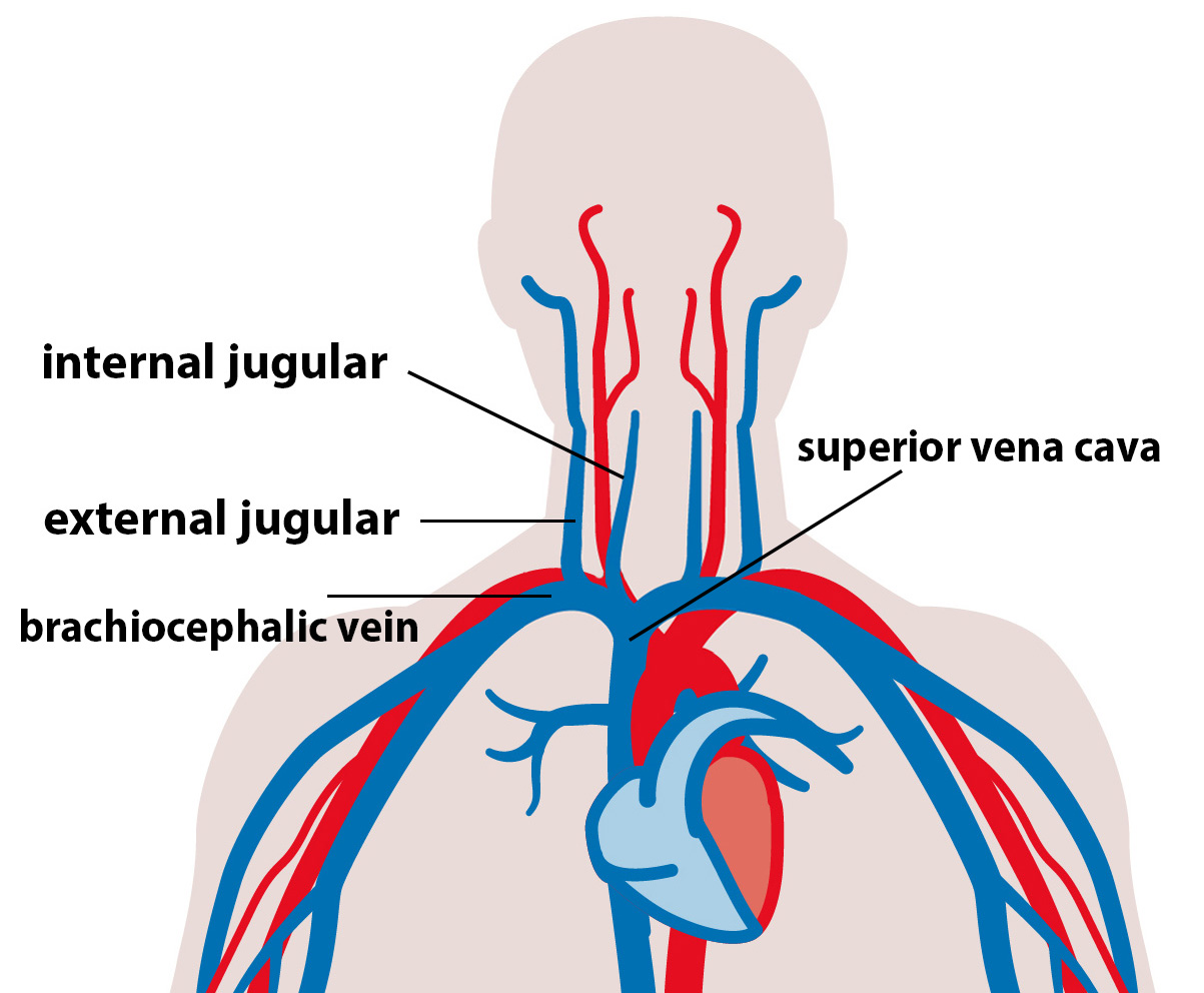
We spoke about the internal and the external jugular veins. The deoxygenated blood in the external jugular vein then drains into the subclavian vein. Subclavian means under the clavicle– this vein travels under the clavicle.
The internal jugular vein then joins with that subclavian vein to form the brachiocephalic vein. That brachiocephalic vein goes into the superior vena cava which then takes the blood right back to the right atrium of the heart.
What she’s suggesting is that a piece of glass went into maybe the internal jugular vein and then got through that brachiocephalic vein into the superior vena cava, punctured it, and then that is causing blood to leak out into the pericardial sac leading to pericardial effusion.
And, this apparently was what they found. They were able to extract a small shard of glass from the boy’s heart.
Discovering this gave the doctors a clearer approach to managing the patient. They will most likely be repairing a hole in the damaged wall and draining the blood from inside the pericardial sac. After all of these are done, it will allow the heart to expand again and continue its normal function.
Summary
- A reduced amplitude as reflected in a patient’s ECG implies that there are weaker signals coming from the heart which can also mean that the heart is not functioning normally.
- An echocardiogram (ECG) is an ultrasound of the heart that helps in visualizing the organ to see if there are any structural abnormalities.
- Pericardial effusion (based from this episode) occurred when blood flowed into the pericardium due to a trauma in the heart. Note that there could be other causes that may trigger such events. Can you name some of them by leaving a comment below?