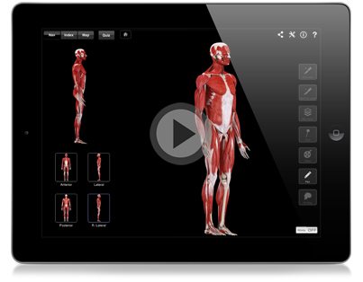
In this video, Leslie talks more about the different superficial muscles of the Anterior Compartment of the forearm. You’ll also learn more about their origins, insertions, actions, and innervations at the end of the transcript.
Enjoy!
Transcript of Today’s Episode
Hello and welcome to another episode of Interactive-Biology TV where we’re making Biology fun. My name is Leslie Samuel and this video is brought to you by our sponsors over at 3D4Medical.com, the creator of this app and a number of other Anatomy apps.

This one is called The Muscle System Pro and it’s available on iPad, iPhone and also for the Mac. You can check them out in the App store.
In this video, I’m going to be talking about The Superficial Muscles Of The Anterior Compartment. More specifically, I’m going to talk about the function of the muscles in the anterior compartment. Then, I’m going to talk about the innervation for the muscles in the anterior compartment. Lastly, I’m going to talk about the superficial muscles that you find in that anterior compartment. So, let’s get right into it.
Right here, I’m looking at my Anatomy guy and I’m going to zoom in to the forearm region, of course, since that’s the region I’m talking about.
First, what is the function of the muscles? In order to show you that, I’m actually going to remove some layers. I’m dealing with just the muscles in the forearm superficial muscles so, I’m going to remove the bicipital aponeurosis and also the brachioradialis muscle. Those are gone now so, we can just focus on those muscles.
When it comes to the function of these muscles, let me select one, I am going to show you the function because that’s the best way to do it. These muscles are going to function in flexion of the wrist and also flexion of the digits. Right now, we’re looking at flexion of the wrist.
Another function of these muscles, let me select a different one because that’s the one that’s going to show it to you, and that is going to be pronation of the forearm. You can see I’m pronating the forearm right now in this example.
So, pronation of the forearm and flexion of the wrist and the individual digits. That’s the function.
What is going to be the innervation?
If you remember from the previous video, we spoke about a nerve that comes down through the cubital fossa. It’s not shown in here because this just deals with the muscle but, you have this nerve that’s coming down through the cubital fossa and that is your median nerve. That median nerve is going to get most of the muscles in the anterior compartment with a few exceptions.
Let’s talk about the exceptions now. We’re dealing with the superficial muscles in this video and all of the superficial muscles get innervated by that median nerve except for one. That’s going to be your flexor carpi ulnaris muscle which is this guy here.
With a name like flexor carpi ulnaris, you can imagine that that’s going to be innervated by the ulnar nerve. So, all of the other superficial muscles, the other four superficial muscles are going to be innervated by the median nerve.
All right, so now that we know that, what are these muscles?
These muscles are, the way I want you to remember the sequence going from lateral to medial is:
P.F.P.F.
F
We’ll look at that in a second. They are all coming from a similar region here on the medial epicondyle for the most part, and that is going to be your common flexor tendon, the CFT and that’s coming there from the medial epicondyle.
The first muscle coming off of that common flexor tendon is going to be your pronator teres. That’s the first “P,” pronator teres. The second is going to be your flexor carpi radialis. That’s the first “F.” The third is going to be your palmaris longus and then, the fourth is going to be the one that we just spoke about, the flexor carpi ulnaris. That’s the P.F. and the P.F.
I want to talk a little bit more about this. In terms of remembering the F’s, the first F was the flexor carpi radialis. This is the lateral side, and this is the medial side. Actually, I should turn it a little like that and once again, this is lateral and this is medial.
The lateral side is where you have your thumb and also, where you have your radius bone, the medial side is where you have another bone and that is the ulnar bone. So, the radius and ulna.
Now, with Fs, it’s flexor carpi for all of them. “Flexor, “that’s the function. “Carpi” refers to the hand, and you either have “radialis” whether it’s on the radial side, the side of the radius bone and then, on the side of the ulnar bone which is going to be more medial, you’re going to have the flexor carpi ulnaris. That’s how you remember the Fs.
In terms of the Ps, the first one is going to be your pronator teres. That’s the one that wraps around and goes laterally so that, when it contracts, it pronates the forearm. The second one is the palmaris longus and the nice way to remember that is you have this, starting right here, this really long tendon that’s going down into the region of the palm. That’s your palmaris longus.
Once again, for the first four muscles, that’s going to be P, which is your pronator teres; F, flexor carpi radialis; P, palmaris longus and then, F, flexor carpi ulnaris.
Then, we have one last muscle and that is your flexor digitorum superficialis. That is this muscle that you’re seeing under here that goes under the other muscles. All of these is flexor digitorum superficialis.
When you’re looking in a cadaver for example, what you’re going to see is that you have multiple tendons that are coming off of that flexor digitorum superficialis. You’ll see the split-tendon like nature of that flexor digitorum superficialis as it goes into the region of the hand to insert on some of the digits.
All right, so in review of what we’ve looked at so far. In terms of the functions of the anterior compartment, we have flexion of the wrist and digits and pronation of the forearm.
In terms of the innervation for the superficial group, they are all innervated by the median nerve except for flexor carpi ulnaris which is innervated by the ulnar nerve.
In terms of the muscles, we’re going to have the P.F.P.F., which is going from lateral to medial, pronator teres, flexor carpi ulnaris, palmaris longus, flexor carpi radialis with our flexor digitorum superficialis.
That’s pretty much it for this video. I hope you got tons of value from it. If you want more information on the origins and insertions, innervations, and actions of these muscles, specifically, this is Episode 105 so, go to Interactive-Biology.com/105 and you’ll get the resources specifically dealing with this video.
So, that’s pretty much it for this video. This is Leslie Samuel from Interactive Biology TV.
That’s it for this video and I’ll see you on the next one.
[table “” not found /]
Thank you a lot. I’m from Czech republic and although the videos are in english I understand it very well. You helped me a lot.
Good video !!! In the review at the end you incorrect name the Flexor carpi Radialis m. and Flexor carpi Ulnaris m.” at 8:10 – 8:16.
I agree with Jesica, I noticed it too, but its okay you repeated it ever so often. You may just make a note towards the end to straighten that out. Over all good work!!!
U r a life saver
Is there a video on the posterior compartment of the forearm??
Great video, but i think you mixed up flexor carpi radialis and ulnaris on the summation. Just thought you oughta to know 🙂