The first bone of the arm and forearm to be discussed in this series is the distal humerus. Its structures including the capitulum and trochlea, the medial and lateral epicondyle, the medial and lateral supracondylar ridges, and the olecranon, radial and coronoid fossae will be discussed by Leslie in this video.
Enjoy!
Transcript of Today’s Episode
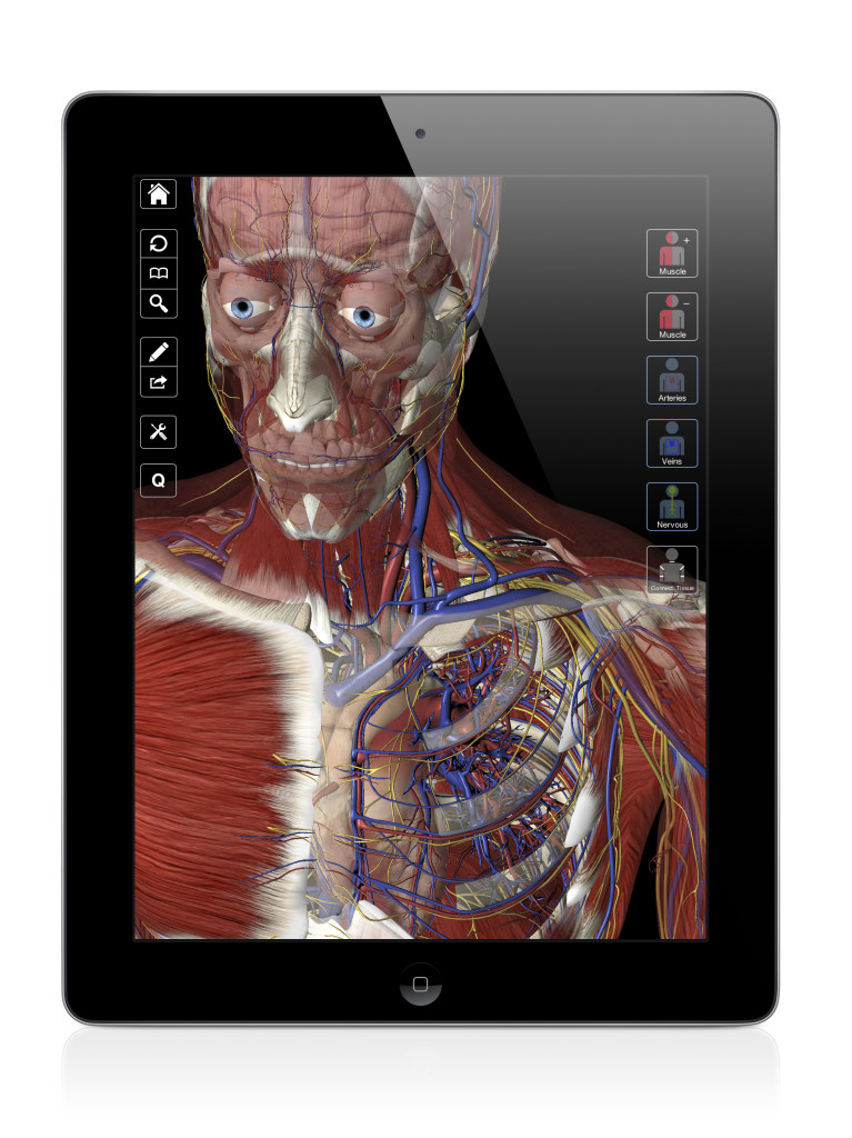
Hello and welcome to another episode of Interactive Biology TV where we’re making Biology fun. My name is Leslie Samuel and this video is brought to you by our sponsors over at 3D4Medical.com, the creators of this app and many other 3D Anatomy app, specifically for the iPad. You can check them out in the App store. This one is called Essential Anatomy. It’s pretty sweet and you’ll see it as I record this video.
In this video, I’m going to be talking about the structures on the distal humerus. We’re beginning to talk about the bones of the arm and the forearm. The first bone of the arm that we want to look at is the humerus. That’s this guy right here. So, let’s look at that. We can fade the others and pay attention to that.
But, what we’re going to do is we’re going to look at the distal humerus because we looked at the proximal humerus when we were talking about the shoulder. Now, that we’re into the arm and forearm, we’re going to be looking at the distal part of the humerus and the structures that we find there.
So, what are the structures that we find on the distal end of the humerus?
The Capitulum and Trochlea
The first one I’m going to look at, keep in mind that this is medial and this is lateral. So, the first structure we’re going to look at is called the trochlea. The trochlea is this structure here that we can call also the medical condyle.
Trochlea comes from a Latin word that means pulley and if you look at the structure of that trochlea, it kind of looks like a pulley that you’d be using to move certain objects at a certain angle and all that fun stuff. That here, that looks like a pulley looks kind of like a spool actually, a spool of thread also, that is called the trochlea and that is going to be your medial condyle.
Then, just lateral to that, we have another structure that’s called the capitulum (I like that word), “capitulum.” That’s also called the lateral condyle. Capitulum, that comes from a Latin word that means a little head. It kind of looks like this little round head right here on the lateral aspect of that distal humerus.
So, first, we have our capitulum that’s lateral and we have medially our trochlea.
Medial and Lateral Epicondyle
Then, we have the epicondyle. We spoke about the lateral and medial condyle. Now, we’re going to talk about the epicondyle. When you hear that prefix, “epi,” that means upon. So, “right upon” or “right here,” you’ll see we have our medial epicondyle and our lateral epicondyle. (Maybe we have to turn that a little bit to see the lateral. Yes, that looks a little better.)
So, this is our lateral epicondyle right here, and this structure medially is our medial epicondyle. If you take your fingers and you palpate the medial aspect of your elbow, you’ll feel this big ball. That’s your medial epicondyle. That’s this guy that you see right here. You can see it significantly larger than the lateral epicondyle.
And, if you are to palpate the lateral aspect right there by your elbow, you won’t feel as big a bump as if do it medially. So, that’s our medial and lateral epicondyles.
Medial and Lateral Supracondylar Ridges
Then, just above those, we’re going to have our lateral and medial supracondylar ridges. This is our medial supracondylar ridge and this is our lateral supracondylar ridge. And, a ridge is exactly what it sounds like. You can see it if I look at it from the side. It looks a little sharper that way. This is your medial supracondylar ridge.
Remember, epicondyle, right upon the condyles and then, we have our supracondylar ridges that are projecting up away from the epicondyles.
Coronoid, Radial and Olecranon Fossae
Then, on the anterior surface, we have a little fossa. Anytime you hear fossa, it comes from the Latin word that means a ditch. So, on the anterior aspect, we have our coronoid fossa which is this guy right here. Why do we call it the coronoid fossa?
Well, right here on the ulna, we have a structure that we call the coronoid process. When you flex your elbow, what’s going to happen is that coronoid process is going to go around the trochlea and fit right here into the coronoid fossa.
Then, lateral to that we’re going to have another little ditch, another little fossa that we call the radial fossa. That’s right above the capitulum. The reason we call it the radial fossa is because right here you have the head of the radius. When you go into elbow flexion once again, the head of the radius is going to go around the capitulum and there’s this groove for it right there that’s called the radial fossa.
One more structure we need to know on the distal humerus, and to do that we’re going to turn this guy around and we’re actually going to look at it on the opposite side. That is another fossa on the posterior aspect. That is going to be a little larger. It’s triangular shaped. That is going to be your olecranon fossa. The reason we call it the olecranon fossa is because when you extend your elbow or you extend you arm, we have this structure on the the ulna that’s called the olecranon process. That’s going to fit right there into that groove that we call the olecranon fossa.
So, let’s review really quick. We have our, and we’re going to start from the back since we are already at this view. This structure is called the olecranon fossa. Then, we turn him around and go to the anterior aspect and we’re going to have medially, we have our trochlea. Laterally, we have our capitulum, then, we have our medial epicondyle and lateral epicondyle. We have our medial supracondylar ridge and lateral supracondylar ridge. We have our coronoid fossa and we have our medial fossa.
That’s pretty much it for this video. I hope you enjoyed it and got some value from it. Of course, if you want more videos like this and other resources to help make Biology fun, head on over to the website, interactive-biology.com.
This is Leslie Samuel. That’s it for this video and I’ll see you in the next one.
are you now an anatomy teacher?
you are the best! wish there were lower limb videos for my anatomy test in 3 weeks !!! these videos are saving my life ha thanks so much
you saved my MD degree, what drug would you like for life?
Not sure how far I will reach in 5 videos, but I don’t think I’ll be quite there yet unfortunately. But we’ll see 🙂
Yes I am indeed. Last semester was my first time teaching anatomy at my new job.
Haha, sorry – no drugs for me, but I’m glad to help.
please continue your passion in biology
i love it how your voice sounded so excited when you mentioned the names. it made me excited too 😀
This is awesome! I’ve been following you ever since I’m an IB student. Now I’m doing MBBS and your videos have helped me throughout my student life. Thank you so much for your biology as well as anatomy teachings 😀
Even though I have to study this in German this is very, very helpful. Many thanks!
What is the software you are using? Thanks!