Today, Let’s talk about another popular topic: Spinal reflexes.
As humans, we are capable of a wide array of conscious movements, and conscious control of our behavior. Much so to the point that we often consider ourselves “conscious animals” to the point of forgetting that a good portion, in fact most of our behavior and actions, are controlled by unconscious reflexes.
The most famous spinal reflex is surely the myotatic (stretch) reflex as it is the most commonly studied reflex in a classroom setting.
The Myotatic Reflex
If the name “myotatic reflex” doesn’t ring a bell, this is the reflex that your physician checks on when he sharply taps the tendon in your knee, and you end up moving your leg as a result.

This reflex involves only 2 neurons, one synapse, one stimulus, one muscle, and one response.
Here is what happens:
[unordered_list style=”arrow”]
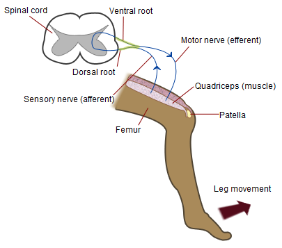
- When the physician taps the tendon in your knee, the pressure from the tap effectively “stretches” the tendon at this point. If you remember well, we saw that tendons contain stretch receptors (muscle spindle and neurotendinous organs) that fire up an action potential when the receptor’s membrane is being stretched. in our example, this “stretch” becomes the stimulus.
- The action potential (nerve impulse) then travels up the leg all the way to the spinal cord throughout an afferent neuron (afferent = going towards the Central Nervous System). It enters the spinal cord at the dorsal horn and ends up synapsing on another neuron in the frontal horn of the spinal cord. As you can see on the diagram, the neuron does not cross over to the other side of the spinal cord. It stays on the same side (if it entered the spinal cord on the left side, it stays on the left side). In other words, it stays ipsilateral. This afferent neuron is the first neuron involved in this reflex.
- In the frontal horn, it synapse on an efferent (efferent = moving away from the CNS) motor neuron. This motor neuron is the second and last neuron involved in this reflex. The motor neuron then, in turn, sends an impulse (action potential) down the leg all the way back to the muscle that has just been stretched.
- At the end of this motor neuron you will find a neuromuscular junction. The neuromuscular junction is what translates/converts every nerve impulse into a muscle contraction. This muscle contraction is what ends up moving your leg almost instantaneously. This muscle contraction is also the “response,” the end product of the reflex.

As a final note, and to be accurate, the muscle involved in the myotatic reflex is the quadriceps femoris muscle.
What Is The Purpose Of The Myotatic Reflex?
Now that we’ve looked at the mechanism behind the knee jerk reflex, let’s talk a little about why this reflex involves only one muscle (this tends to be a property of every stretch reflex).
You might find it interesting to think of it in these terms: every unconscious / involuntary stretch of your tendon is probably not something that you wanted in the first place. As a response it seems totally fair to have a system in place that automatically attempts to restore the contraction of your muscle the way you intended it to be.
Another way to view this is to look at how we stand. In order to stand, we need every muscle in our body to be contracted exactly the right amount. Any change in that will make us loose our balance and fall. When it comes to the legs, it makes perfect sense that we would have a system in place whose purpose is to prevent us from falling when, for some reason that we did not choose, something happens to stretch one of our muscles. It is very important to re-contract that muscle to the right amount as soon as possible!
Can you think up any other purpose this reflex might have?
Minimum To Remember
The myotatic reflex involves:
[unordered_list style=”tick”]
- One Muscle: The quadriceps femurs muscle
- One Stimulus: Stretch of the tendon
- One Stimulus: Stretch of the tendon
- One synapse: In the ventral horn of the spinal cord
- Two neurons:
- [ordered_list style=”decimal”]
- One afferent that goes from the leg to the spinal cord
- One efferent (motor) that goes from the spinal cord back to the muscle.
[/ordered_list]
- One response: Muscle contraction
If you want more articles and videos about the Nervous System, you can find them here. More resources are available to help make Biology fun. I invite you to absorb all the content you can find here at Interactive-Biology.com.
