Now that we’ve covered the external structure of the coolest cell in the body–the neuron(!), let’s look at its internal structure.
Smaller Structures Inside The Neuron
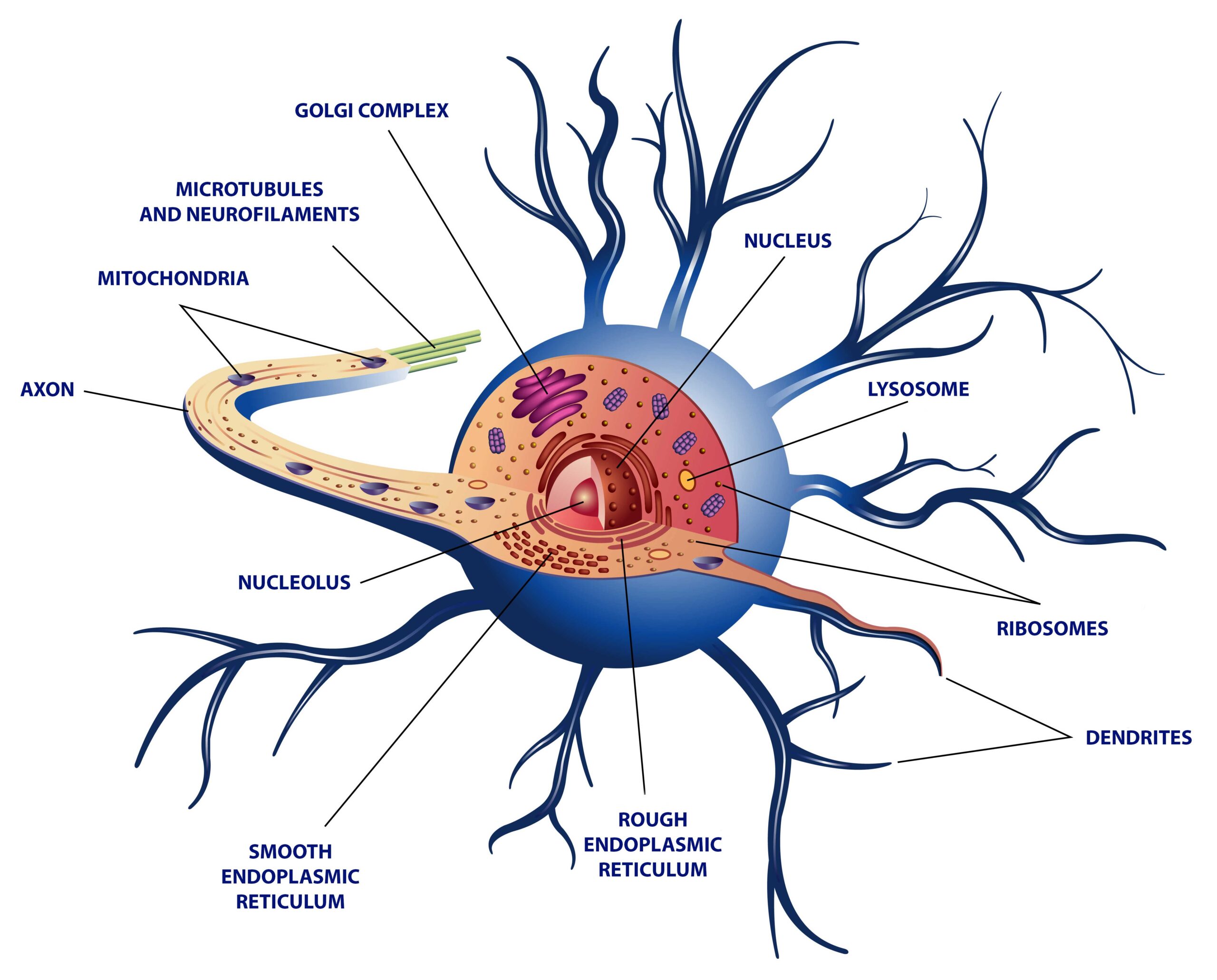
Like most cells, the neuron contains:
- Nucleus
- Mitochondria
- Golgi bodies
- Endoplasmic reticulum and Ribosomes
- Transport Channels – microtubules, neurofilaments, and neurotubules
Nucleus
The nucleus contains the chromatin and the genetic make-up of the organism. The nucleus is generally located in the soma of the cell.
But, did you know that the nucleus in a neuron can “travel?”
- Like we said, the nucleus is generally located in the soma, but if the neuron gets injured, the nucleus will be found at the extremity of the cell. (An eccentrically located nucleus is a pathological sign of injured nerve tissue).
Mitochondria
… And, a lot of it. The neuron is a very active cell that requires lots of energy
Did you know that your brain consumes 20% of your calorie intake… just for thinking!
Mitochondria can be found anywhere in the neuron but are mostly concentrated in the cell body and in the synaptic terminals.
Golgi bodies
The Golgi bodies package proteins synthesized by the cell.
Endoplasmic Reticulum and Ribosomes
The Endoplasmic reticulum and ribosomes synthesize proteins.
Transport Channels
Here is another really cool characteristic about the neuron: protein synthesis can occur far from the nucleus! In order to increase synaptic growth and the speed of information processing, so that you may learn and remember things faster, neurons can synthesize new proteins directly at the extremities 🙂
Sometimes, however, proteins cannot be made on the spot and they need to be transported from one part of the cell to a different part of the cell. In this case, the proteins can travel along “transport channels” such as microtubules, neurofilaments, and neurotubules.
Signal Alchemy In Chemical Synapses
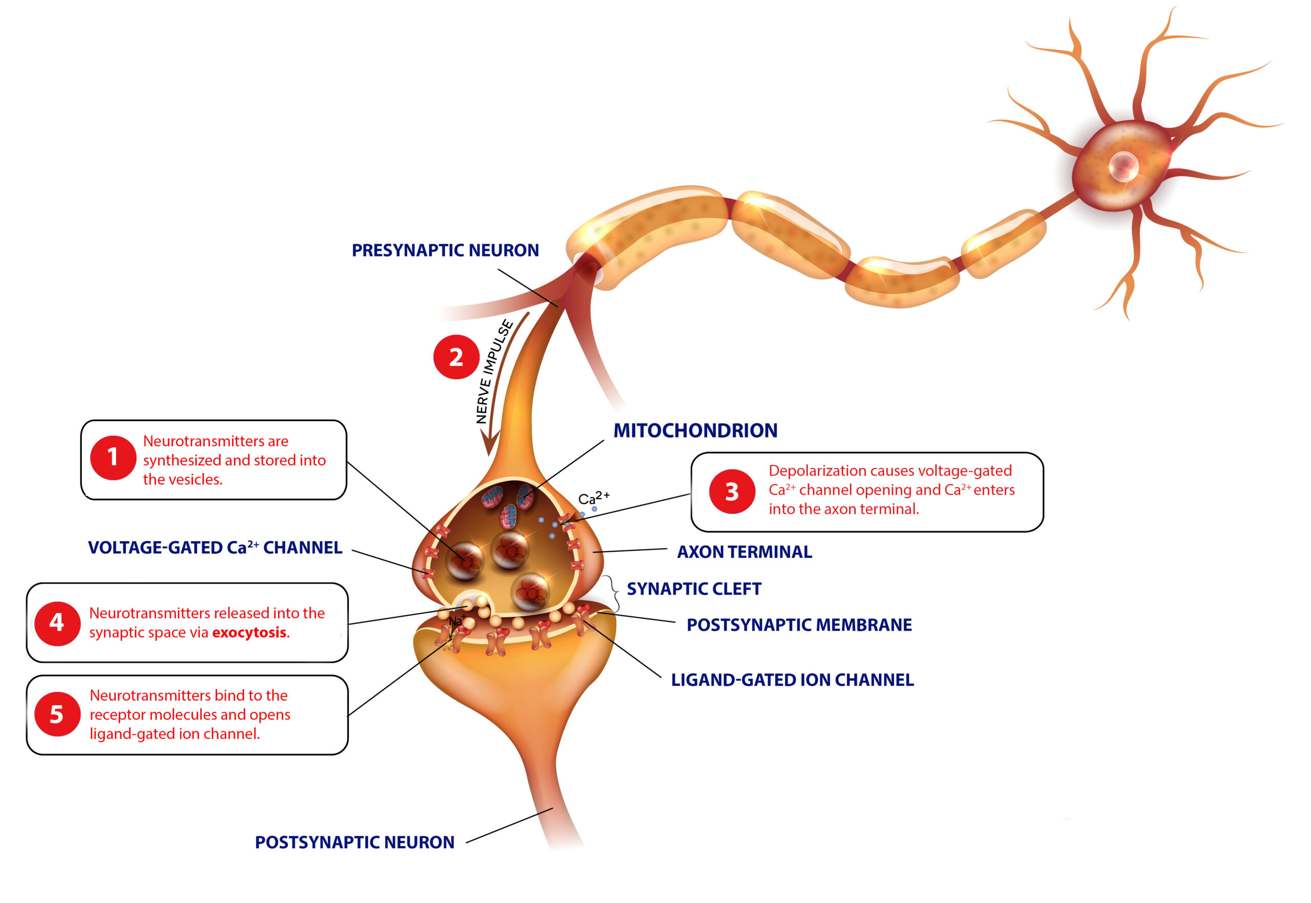
Zoom-In On The Presynaptic Terminal
The presynaptic terminal, at the end of the axon, is a very busy and active area.
- This is where the electric signal from the axon is transformed into a chemical signal (neurotransmitter) that can bridge the gap between two adjacent neurons.
The place is filled with mitochondria for energy, neurotransmitters in vesicles ready for release into the synaptic cleft, neurotransmitter fragments that have been endocytosed for recycling, and a plethora of other proteins and structures that would require a book to describe!
At this level, the cell membrane is filled with different types of ion transporters.
- Every time a signal comes down from the axon, the electric pulse alters the polarity of the cell membrane. This forces ions (calcium–for example) to rush into the cell, other ions will rush out of the cell, and the whole thing starts a fuss of activity resulting in the release of neurotransmitters in the synaptic cleft.
Zoom-In On The Postsynaptic Structure (at the dendrite)
- This is where the neuron receives the chemical information from the previous cell’s presynaptic terminal, and transforms it into a new electrical signal.
On this side of the synaptic cleft, the membrane is filled with different types of receptors (ionic and metabotropic receptors) that receive free neurotransmitters.
In some synapses, right below the membrane is a region called the postsynaptic density. It is called like this because it looks really “dense” on a microscope due to the number of proteins that are present at this level.
Electrical Synapses – The Other Kind of Synapses
Did you know that the human body also contains “electrical synapses?” These synapses do not require neurotransmitters.
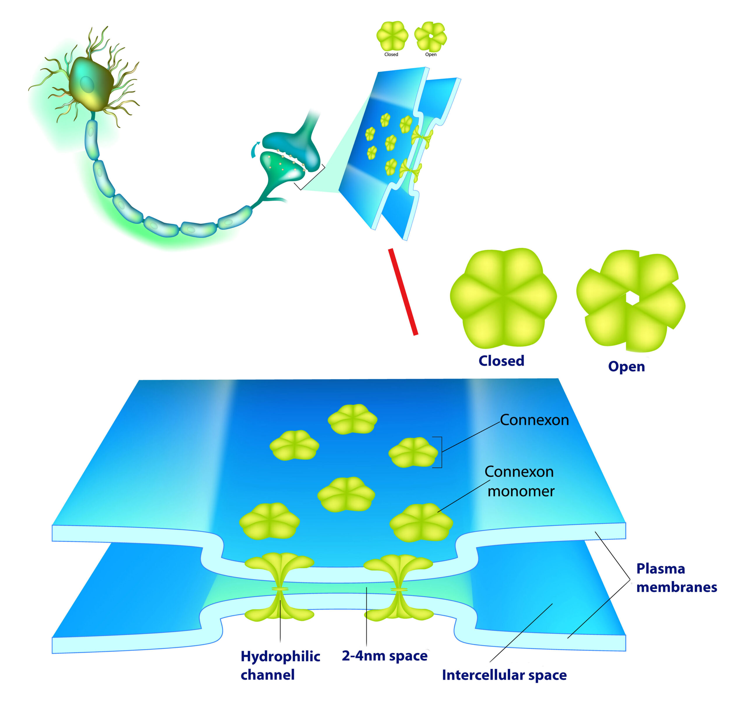
In electrical synapses, the two cells are so close to each other that the electrical pulse can “jump” over the gap – the gap junction.
To keep the two cells close together, the membranes on both sides house protein structures called connexons. These channels can cross both membranes and are large enough to allow the diffusion of ions (i.e. the electrical signal) between these two cells.
- I invite you to read more about synapses and synaptic activity if you are totally enthralled by the magic of the synapse!
That’s it for this introduction to the neuron’s internal structure.
If you want more articles and videos about the Nervous System, you can find them here. More resources are available to help make Biology fun. I invite you to absorb all the content you can find here at Interactive-Biology.com.