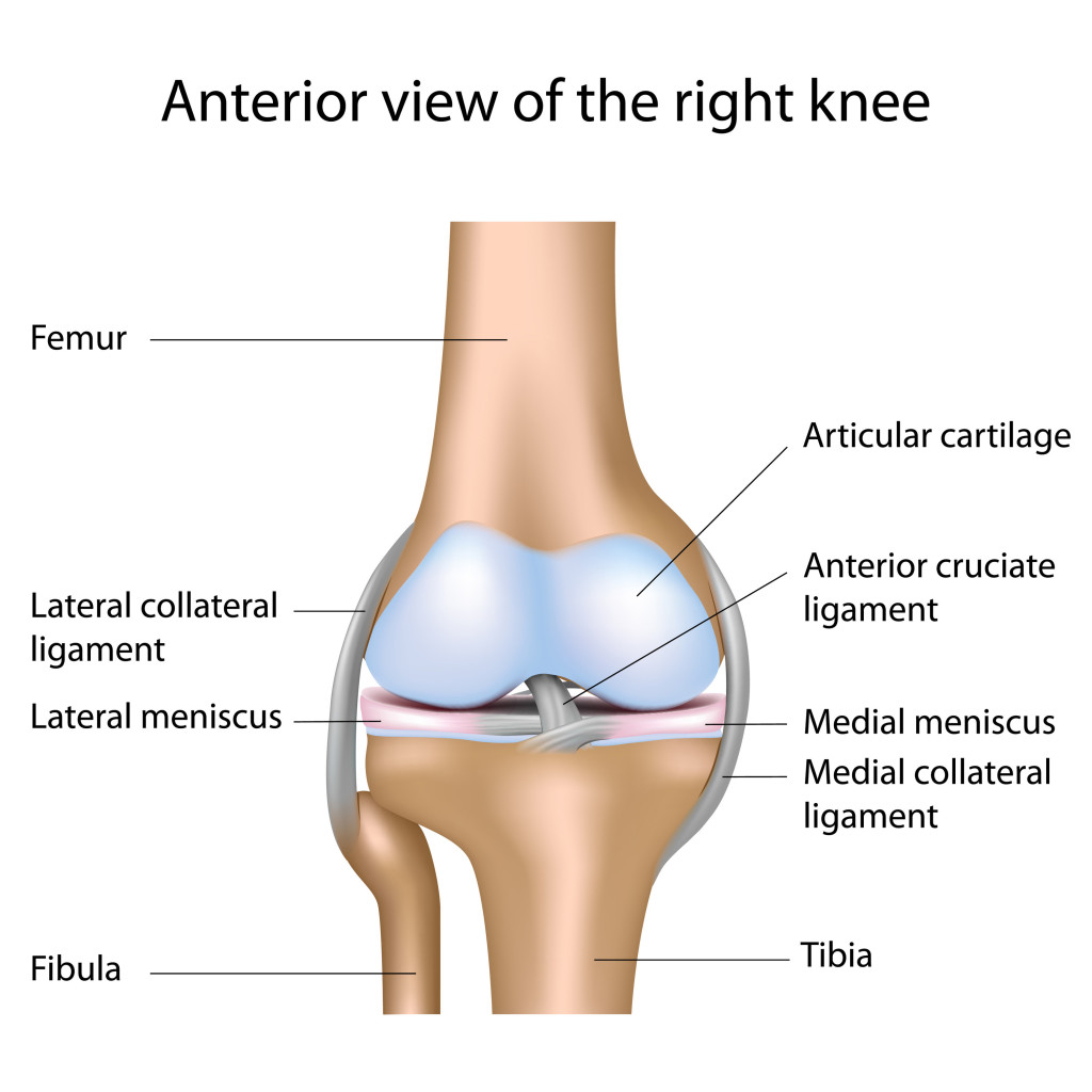
The knee is a joint formed, stabilized, and given mobility by the articulation of bones, ligaments and tendons. This joint is the largest joint in the body and is formed by the articulation of the femur bone in the thigh with the tibia in the lower leg.
What Type of Joint is a Knee Joint?
There are 3 main types of joints:
- Fibrous – an immovable joint;
- Cartilaginous – partially moveable; and
- Synovial – a freely moveable joint.
The knee joint is classified as a synovial joint for obvious reasons based on the definitions given.
Synovial joints or diarthroses are considered moveable joints. According to the Encyclopedia of Nursing and Allied Health, joint function, synovial joints can be further divided into 3 types. These are:
- Uniaxial – joints that hinge or pivot moving only in one plane;
- Biaxial – such as the saddle and condyloid joints. These joints move in two planes. And lastly,
- the Triaxial – which allows movement in three planes including the ball and socket joints and gliding joints.
The knee falls under the uniaxial as it is a hinge joint and it moves in one plane with slight rotational movement, but the rotation is not enough to be considered significant.
Anatomy and Physiology of the Knee
The knee is the largest joint of the body and is often the site of pain and injury in athletes (the reason I am writing about this is that I strained my medial collateral ligament, more on that later). It consists of the medial and lateral condyles (round projection at the end of the bone) of the lower femur (thigh bone) and the same condyles at the upper end of the tibia.
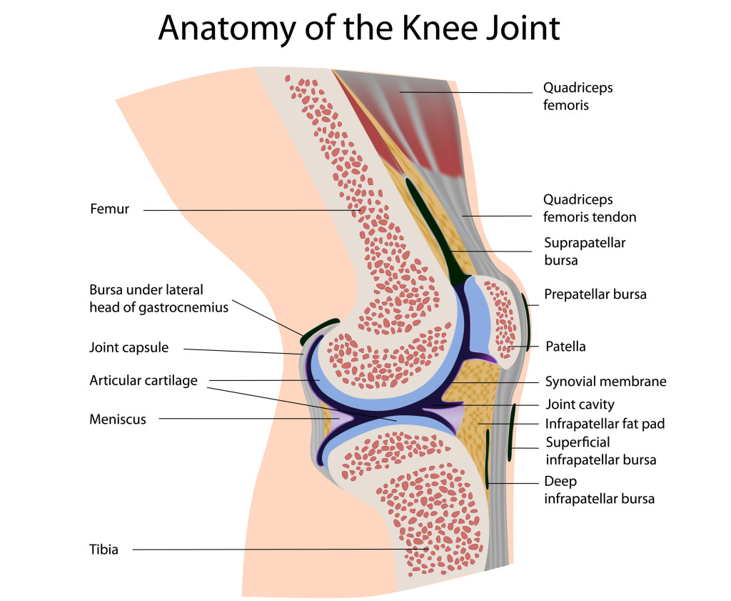
The patella (knee cap) covers the front of the joint. It is the protruding structure you see when you extend your knee. This structure slides along a groove on the femur.

Each of the 3 bones in the knee joint is covered with articular cartilage, which is a tough elastic material, that acts as shock absorbers and allows the knee joint to move with ease. Another cartilage tissue called the menisci separates the femur and tibia, divided into two crescent-shaped discs located medially and laterally (inner and outer respectively). This cartilage also acts as shock absorbers, as well as enhancing stability.
In a normal knee joint, a synovium (synovial membrane) surrounds the knee joint. It produces synovial fluid that nourishes the surrounding cartilage in the knee. The synovium also functions in protecting and supporting the joint due to its tough outer layer.
Stability of the Knee
The stability of the knee is due mainly to four ligaments. A ligament is several large fibrous bands of tissue, comparable to that of a rope, they support the knee on both sides and front to back. Ligaments connect bone to bone.

The ligaments that connect the femur to the tibia and fibula are as follows:
- Medial Collateral Ligament (MCL) provides stability to the inner (medial) aspect of the knee. It is also known as the Tibial Collateral Ligament because it connects the Femur and Tibia
- Lateral Collateral Ligament (LCL) provides stability to the outer (lateral) aspect of the knee. It is also known as the Fibular Collateral Ligament because it connects the Femur and Fibula
- Anterior Cruciate Ligament (ACL), in the center of the knee, limits rotation and forward movement of the Tibia
- Posterior Cruciate Ligament (PCL), also in the center of the knee, and like the ACL, secondarily limits rotation, while primarily limits backward movement of the Tibia.
Movement of the Knee
Your knee is a hinge joint like your elbow, which we discussed already, which means it bends and straightens. We also said that it has the ability to slightly rotate as it moves.
The muscles in the thigh, the quadriceps and hamstrings, perform movement of the knee joint, but these muscles need assistance from tendons to connect them to the muscles. Tendons are tough cords of tissue that connect muscle to bone. They are similar to ligaments in structure. The difference is in just what they articulate with.
When you straighten your leg, the quadriceps muscles contract pulling on the quadriceps tendon, which in turn pulls on the patella via the patellar tendon causing an extension of the knee. Please note the patellar tendon connects the patella to the tibia. So, technically, it is a ligament but commonly called a tendon.
On the posterior side of the knee, the hamstring group of muscles contract pulling on the tendons associated with the hamstring, pulling on the femur, which in turn causes the flexion of the knee.
Conclusion
- The knee is a type of synovial hinge joint.
- The knee is formed by the articulation of the femur bone in the thigh with the tibia in the lower leg.
- A patella or knee cap covers the front of the joint.
- A synovium surrounding the knee joint produces synovial fluid that nourishes, protects, and supports the joint.
- Four types of ligament supports the knee on both sides and front to back. These are the Medial Collateral Ligament (MCL), Lateral Collateral Ligament (LCL), Anterior Cruciate Ligament (ACL), and Posterior Cruciate Ligament (PCL).
- Muscles involved in the movement of the knee joint are the quadriceps and hamstring group of muscles with the help of some tendons.

Thanks for this informative overview.
thanks for the information provided