In my last article, I discussed the types of bones and how they were formed. I left out quite a bit on joints because their classification and structure results in a lengthy article, too.
Joints are just as important as our muscles when it comes to movement. A joint is a point where two bones meet and their purpose is to allow movement, but there are some exceptions. Joints are classified in two major ways: functional and connection.
Joint Classification: Functional (Movement)
 The first type describes a joint where no movement occurs, such as the joints of our skulls. These can also be thought of as sutures. Synarthrosis is the term used to describe this type of joint.
 The next type of joint is capable of movement, but it is limited. Our vertebrae are a classic example of this type of joint, which is called an amphidiarthrosis.
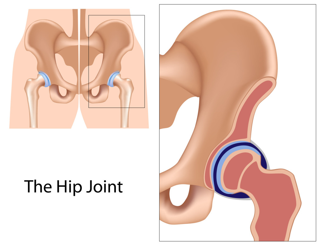
Our free moving joints, such as the knee and hip, are classified as diarthroses.
These terms can be a bit complicated to remember, but if you break down the words and the meaning, it is much simpler. I have used this method a countless number of times and it helps me.
For instance, the root word is arthrosis and means joint. The prefixes of each term differentiate the words from each other. “Syn” means fusion, which makes sense because the bones of the skull are fused together and cannot move. “Amphi” can mean “both”. Amphidiathrosis can be thought of as a mix of synathrosis and diarhthrosis. “Di” means two, and diarthroses have a cavity between two articular surfaces. This will make more sense in a few paragraphs.
Joint Classification: Connection and Structure
Each of the following classifications is related to the previous classifications. Fibrous joints cannot move for the most part, so they are called synarthrosis joint or an amphidiarthrosis joint. Fibrous joints also have a few sub-classifications: gomphoses, sutures and syndesmoses.
Gomphoses are the joints that connect the teeth to the sockets of the maxilla. Sutures are the joints of the skull and syndesmoses are found between the long bones of the arm and leg. Unlike other fibrous joints, syndesmoses are capable of movement, but not to the extent of diarthroses.
Fibrous joints are called such because they are connected by dense connective tissue, usually collagen. Fibrous joints also lack a joint cavity.
Cartilaginous joints allow for more movement than fibrous joints, but less movement than synovial (free moving) joints. Rather than being connected by collagen, they are connected entirely by cartilage. Two types of cartilaginous joints: primary (sychondrosis) and secondary (symphases) exist.
Primary cartilaginous joints are only joined by hyaline cartilage while secondary cartilaginous joints are connected by hyaline and fibrocartilage. Like fibrous joints, cartilaginous joints lack a joint cavity.
Synovial joints are the ones we are most familiar with and they are the most common in the body. Joints such as those in the knee, hip, wrist and shoulder are classified as synovial joints because they are capable of moving freely. Synovial joints can move in a variety of different ways. They can rotate, flex (bend), extend (straighten), abduct (move away from body’s mid-line) and adduct (move towards the body’s midline). There are many types of synovial joints that are named based on their shape. The types of synovial joints are:
1.) Hinge. Our knees represent a hinge joint. This one is easy to remember because they work just like a door hinge. This is also easy to see when you let your legs dangle over the side of a pool or chair, and when your doctor checks your reflexes. Hinge joints do no not have a full range of motion and can be extended or flexed.

2.) Gliding. This type of synovial joint is found in the wrists and they slide on top of each other. I always think of two blocks, one on top of the other, moving back and forth.
3.) Saddle. Saddle joints look just like, well, a saddle. Like gliding joints, saddle joints are also found in the wrist. They allow a wide variety of movement including flexing, extending, abduction and adduction.
4.) Condyloid. This type is unique because of its shape, which resembles an ellipsis. The bones of this joint fit into one another. Like saddle joints, they have a wide variety of movement. The only exception is rotation. Our lower jaw is an example of a condyloid joint.
5.) Ball and Socket. Ball and socket joints include the shoulder and hips. This type allows for the most movement and is the most flexible.

6.) Pivot. This is the final type of synovial joint. With pivot joints, one bone rotates around another and they are not capable of any other kind of movement. An example of a pivot joint would be the radioulnar joint and the joint between the C1 and C2 vertebrae.
Synovial Joint Structure
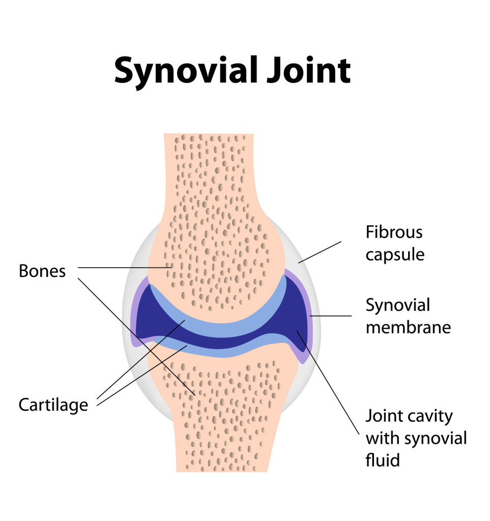
The free movement of synovial joints is not the only quality that separates them from the rest. They also have a unique structure. There are three structures that all synovial joints have: the articular capsule, articular surface and synovial cavity.

Synovial joints are surrounded by the fibrous articular capsule. This capsule is avascular (no blood vessels) but it is innervated, so it is sensitive to movement. Hyaline cartilage surrounds the articular surface, which covers the bones of the joint. It is avascular and it is not innervated. Synovial fluid lubricates the cartilage in order for the joints to move freely. The synovial cavity is located between the articular surfaces and it holds the synovial fluid.
Joint Stability
A joint is only useful if it can remain stable. Joint stability refers to how resilient the joint is against stress. There are three major ways a joint can be stabilized.
The first way of stabilizing a joint is their shape. Each type of joint is designed for a specific motion and the shape helps the resiliency of that joint. Ligaments are also important for joint stability. They guard against excess stress and movement. Muscle tone is also an important factor in stabilizing joints. Muscles surrounding the joints prevent them from collapsing.
An example of how muscle tone affects stability can be found in the arches of our feet. The bones in our feet are small and the arches would easily collapse without the muscles.
Our joints are every bit as important as our muscles when it comes to movement and more complex than one might originally think. Importance of joints often goes unnoticed until something goes wrong. Without joints that function properly, movement would be impossible.
