If you’re a neuroscience student, you’ve got to love or hate the thalamus!
Why? Because it will come back over and over and over again in all your neuroscience, neuroanatomy, and biopsychology classes. It is so important and so complex that professors love to test students on it.
So, there you have it. If you really spend the time to study the thalamus and understand it like the back of your hand, you will love it. It’s your key to beating all your competition and getting those coveted As.
If you can’t seem to wrap your head around it, you will hate it for the rest of your studies, because it will come back to haunt you on almost every midterm!
Let’s get our basics straight.
Thalamus Basic Structure and Terminology
We have a left and a right thalamus. Be aware of that when you are given a slice of the thalamus to identify.
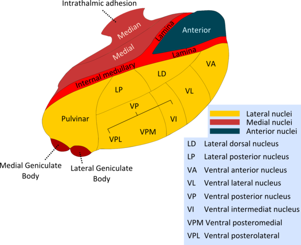
The thalamus is divided along the anterior to posterior axis (front to back) by a thin strip of myelinated fibers called the Internal Medullary Lamina (IML).
At the anterior end, the Internal Medullary Lamina splits into two “branches.” The area in between those branches is called the Anterior nucleus (A).
Way at the posterior end of the thalamus we find the Pulvinar (Pul) nucleus (the pulvinar plays an important role in vision as a relay station in the tecto-fugal pathway).
Under the pulvinar you will find two small nuclei: the Lateral Geniculate body (important for vision in the Retino-geniculo-calcarine pathway), and behind it lies the Medial Geniculate body (important in hearing).
The Internal Medullary Lamina also serves to divide the thalamus into a medial part (closer to the center of the brain) and a lateral part.
So, at the front we have the anterior nucleus. Then, if we travel towards the back of the brain we will find on one side of the IML the DorsoMedial nucleus (DM), and on the other side of the IML we will find the Lateral part of the thalamus.

The Lateral part can be divided into additional sections.
Before we go on with more terminology, I want to invite you to really give your attention to the actual words making up these names; they make sense!
- Ventral refers to the “bottom” part of the thalamus.
- Dorsal refers to the “Top” part of the thalamus.
- Anterior refers to the “front”
- Posterior refers to the “back.”
- Lateral means “on the side.”
OK, here we go!
Starting anteriorly, we have the Ventral Anterior nucleus (VA), followed by the Ventral Lateral nucleus (VL) on top of which lies the smaller Lateral Dorsal nucleus (LD) (remember, “lateral dorsal” means “on the side and on top”).
Behind the VL we find the Ventral Post Lateral nucleus (VPL) (again, “ventral post lateral” means “behind the ventral lateral nucleus”), and behind the LD we find the Lateral Posterior nucleus (LP).
At this level, hidden inside the thalamus, you will find the Ventral Posterior Medial nucleus (VPM).
We will stop here for today.
- Remember, the terminology of the thalamus itself is actually pretty straight forward if you pay attention to what the words mean.
Do This To Master The Basic Anatomy
- Take a piece of paper, and draw the outline of a football. Write down all the nucleus of the thalamus (just the names) on a corner of the page.
- Next, look at a diagram of the thalamus, and grossly draw in the Internal Medullary Lamina.
- Now, just by looking at the names, try to draw in the parts in a way that makes sense according to the meaning of the words. You should be able to guess 90% of the anatomy of the thalamus.
- Look up at a diagram of the thalamus again, and draw in the parts you didn’t know.
- If you are able to do this, you will always be able to reconstruct the basic anatomy of the thalamus.

If you want more articles and videos about the Nervous System, you can find them here. More resources are available to help make Biology fun. I invite you to absorb all the content you can find here at Interactive-Biology.com.

I found this explanation really helpful. specially the part where you explain what ventral dorsal etc meant with respect to the thalamus. Before i kept trying to derive the logic behind the nomenclature but i couldn’t. Good job!