Kidney Anatomical Position
Where are the kidneys located in the body?
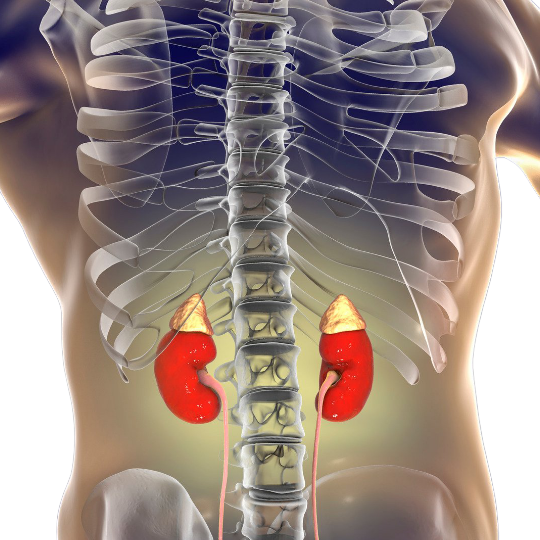
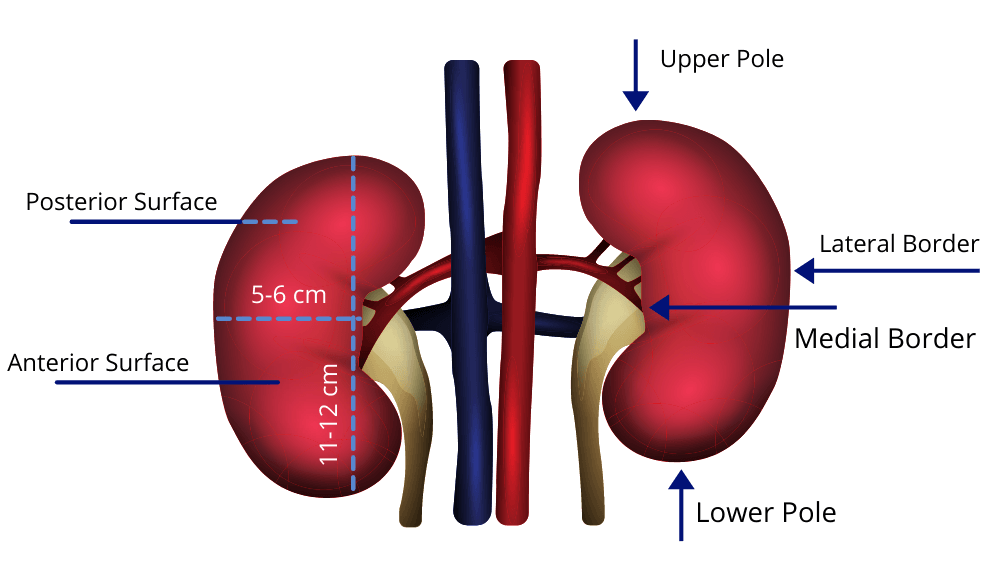
The kidneys are reddish-brown, bean-shaped organs situated retroperitoneal on the posterior abdominal wall. They extend from lumbar vertebra T12-L3. Normally, the kidney is about the size of a mouse and measures approximately 11-12 cm in length, 5-6 cm in width, and 2.5-3 cm in thickness.
Kidney Structure
The kidneys have a superior and inferior pole, medial and lateral margins, and an anterior and posterior surface. The superior pole of each kidney is deep to the rib cage. For the right kidney, its superior pole is at the 12th rib and for the left, the superior pole is at ribs 11 and 12.
On the medial margin of the kidney is a concave region called the renal hilus. The renal hilum is the entrance to the renal sinus. Structures such as the renal veins, artery, nerves, and lymphatic vessels are located in the renal hilum.
The renal sinus is a fat-filled cavity inside the kidney that extends from the hilum. At the hilum, the ureters also exit the kidney. A fibrous renal capsule covers each kidney. They are also surrounded by perirenal fat which extends in the renal pelvis.
Around the perirenal fat, is a layer of renal fascia called the Gerota’s fascia. The Gerota’s fascia has an anterior and posterior layer. The anterior layer continues medially and fuses with the adventitia of the aorta and the inferior vena cava (IVC). The posterior layer fuses with the psoas major fascia.

Internal Structure of the Kidney
If one should take a knife and cut the kidney in halves from the superior pole to the inferior pole, you would find two distinct internal regions of the kidney: the cortex, and the medulla.
The cortex is the superficial outer layer of the kidney located underneath the capsule.
The medulla is the inner layer and it extends from the renal cortex to the renal sinus. The medulla is divided into various cone-shaped structures called the renal pyramids. The renal pyramids extend from the cortex to the renal papilla. These papillae extend into a space called the renal pelvis.

The renal pelvis is a funnel-shaped structure that is continuous with the ureter. The renal pelvis is divided into calyces. Each pelvis receives about 2 major calyces, which in turn receive minor calyx, which in turn collect urine from the papilla.
Posterior and Anterior Anatomical Relations of the Kidney
Due to the location of the kidney, it comes into contact with various structures in the body.
Posteriorly, the kidneys are related to the:
- diaphragm
- quadrates lumborum muscle
- transversus abdominis muscle
- 11th and 12th rib (left kidney)
- 12th rib (right kidney)
- costodiaphragmatic recess
- subcostal, iliohypogastric
- ilioinguinal nerves.
Anteriorly, the right kidney is in relation to the:
Anteriorly, the left kidney is related to the:
Conclusion
We have learned the basic anatomy of the kidney with some of its parts and structures. More of its functions and physiology can be found in this post.
As a supplement to this it is well worth checking out the following pictorial summary that was created by one of my High School Biology Students
http://inspiredscience.squarespace.com/hsc-biology/2012/2/19/kidney-poster-great-pictorial-summary.html
I was looking for more information on how the kidneys spray out blood and how they can be damaged by high blood pressure. I have Type 2 Diabetes and I know they want to keep my blood pressure down for that reason. Also, I think there is input from the kidneys to control blood pressure, but I am not sure of that fact. That is why I came to interactivebiology.com because I heard about the Web site (two existing words, not website – because we do not need a ten-foot thick dictionary to pass on to future generations) on The Tech Guy podcast. I also wanted graphics in that region.
I am creating a presentation for my PhD dissertation and would like to know if I can re-use an image from your site, in particular the image of the structure of a kidney. Thank you, Bianca
what is is minimum size of kidney, is parenchymal changes of grade is treatable
parenchymal changes of grade ii are reversible